

Christos Pliotas, Richard Ward, Emma Branigan
Use of electron paramagnetic resonance (EPR) to understand how the mechanosensitive ion channel proteins, MscS and MscL, gate (open and close) under hypoosmotic stresses (see Fig 1.)
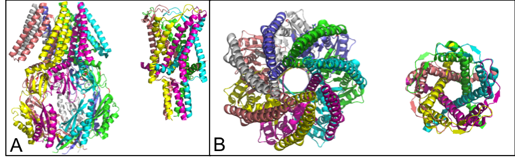
Figure 1: Crystal structures of heptameric MscS and pentameric MscL. A) Comparing the length of MscS (left hand side) to MscL (right hand side). B) Comparing the width of MscS with MscL.
Both MscS and MscL are diamagnetic and therefore invisible to EPR, which detects unpaired electrons. Chemically modifying mutated in single cysteine residues with a label called MTSSL, which contains a stable unpaired electron, overcomes this issue. In addition it means that the label reports on a specific position on the protein. By employing a pulsed EPR experiment called PELDOR it is possible to measure distances (1.5-8 nm) between the monomeric units of these proteins and thus gain structural information (see Fig 2.). Importantly, the protein does not need to be crystalised.
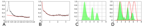
Figure 2: Example PELDOR data for MTSSL labelled MscS D67C mutant. A) Raw experimental data. B) Processed data (black line) and best fit simulated data (red). C) Experimental distance distribution generated from simulated data. D) Using this approach it was possible to show that Mscs is in an open conformation in solution, since a comparison of the experimental distance distribution with one derived from an open crystal structure of MscS gave the best fit [1].
[1] Pliotas C, Ward R, Branigan E, Rasmussen A, Hagelueken G, Huang H, Black SS, Booth IR, Schiemann O, Naismith JH. (2012) Conformational state of the MscS mechanosensitive channel in solution revealed by pulsed electron – electron double resonance ( PELDOR ) spectroscopy. PNAS, 109:E2675-82.